How does my doctor know I have pancreatic cancer?
Q: What are the signs and symptoms of pancreatic cancer?
- Jaundice - Jaundice is a yellowing of the eyes and skin. It occurs in at least half of all people with pancreatic cancer and in all cases of ampullary cancer. Jaundice is caused by the buildup of bilirubin in the body. Bilirubin is a dark yellow -- brown substance that is made in the liver. Normally, the liver excretes bilirubin into bile. Bile goes into the intestines and eventually leaving the body in the stool. When the common bile duct becomes blocked, bile can't reach the intestines, and the level of bilirubin builds up. Cancers that begin in the head of the pancreas are near the common bile duct. These cancers can compress the duct while they are still fairly small. This can lead to jaundice, which may allow these tumors to be found in an early stage. But cancers that begin in the body or tail of the pancreas do not compress the duct until they have spread through the pancreas. By this time, the cancer may have also spread beyond the pancreas. Sometimes, the first sign of jaundice is darkening of the urine from bilirubin. As bilirubin levels in the blood increase, the urine becomes brown in color. If the bile duct is blocked, bile (and bilirubin) can't get through to the bowel. When this happens, a person may notice their stools becoming lighter in color. When bilirubin builds up in the skin, it turns yellow and starts to itch. Cancer is not the most common cause of jaundice. Other causes, such as gallstones, hepatitis, and other liver diseases, are much more common.
- Abdominal or back pain - Pain in the abdomen or back is common in advanced pancreatic cancer. Cancers that start in the body or tail of the pancreas may grow fairly large and start to compress on other nearby organs, causing pain. The cancer may also spread to the nerves surrounding the pancreas, which often causes back pain. The pain may be constant or it may come and go. Of course, pancreatic cancer is not a common cause of pain in the abdomen or back. It is more often caused by a non-cancerous diseases or even another type of cancer.
- Weight loss and poor appetite - Unintended or unexpected weight loss is very common in patients with pancreatic cancer. These people also complain of being very tired and having little or no appetite.
- Digestive problems - If cancer blocks the release of the pancreatic juice into the intestine, a person may not be able to digest fatty foods. The undigested fat may cause stools to be unusually pale, bulky, greasy, and to float in the toilet. The cancer may also wrap around the far end of the stomach and partly block it. This can cause nausea, vomiting, and pain that tends to be worse after eating.
- Gallbladder enlargement - If the cancer blocks the bile duct, bile can build up in the gallbladder, which then becomes enlarged. Sometimes a doctor can feel this enlargement during the physical exam. It can also be detected by imaging studies.
- Blood clots or fatty tissue abnormalities - Sometimes, the first clue that there is a pancreatic cancer is the development of a blood clot in a large vein, often a vein in the leg. This is called a deep venous thrombosis or DVT. Sometimes a clot breaks off and travels to the lungs, making it hard to get enough air. A blood clot in the lungs is called a pulmonary embolism or PE. Still, having a blood clot does not usually mean that you have cancer. Most blood clots are caused by other things. Another clue that there may be pancreatic cancer is the development of uneven texture of the fatty tissue underneath the skin. This is caused by the release of the pancreatic enzymes that digest fat.
- Diabetes - Rarely, exocrine cancers of the pancreas cause diabetes (high blood sugar) because they destroy the insulin-making cells. More often, there are slight problems with sugar metabolism that do not cause symptoms of diabetes but can still be recognized by certain blood tests.
History and physical exam
A thorough medical history will be taken to check for any pancreatic cancer risk factors, and to obtain information about pain (how long it has been present, its severity, its location, and what makes it worse or better), appetite, weight loss, tiredness, and other symptoms. A thorough physical exam will focus mostly on the abdomen to check for any masses or fluid buildup. The skin and the white part of the eyes will be checked for jaundice (yellow color). Cancers that block the bile duct may also cause the gallbladder to become enlarged, which can sometimes be felt on physical exam. Pancreatic cancer may spread to the liver, causing it to enlarge.
The cancer can also spread to lymph nodes above the collarbone and other locations. These areas will be looked at carefully for swelling that might indicate spread of a cancer.
Imaging tests
- Computed tomography (CT, CAT) scan - The CT scan is an x-ray procedure that produces detailed cross-sectional images of your body. Instead of taking one picture, like a standard x-ray, a CT scanner takes many pictures as it rotates around you. A computer then combines these pictures into images that resemble slices of the part of your body being studied.
Before any pictures are taken, you may be asked to drink 1 to 2 pints of a liquid called oral contrast. This helps outline the intestine so that certain areas are not mistaken for tumors. You may also receive an IV (intravenous) line through which a different kind of contrast dye (IV contrast) is injected. This helps better outline structures in your body.
The injection can cause some flushing (redness and warm feeling that may last hours to days). A few people are allergic to the dye and get hives. Rarely, more serious reactions like trouble breathing and low blood pressure can occur. Medicine can be given to prevent and treat allergic reactions. Be sure to tell the doctor if you have ever had a reaction to any contrast material used for x-rays.
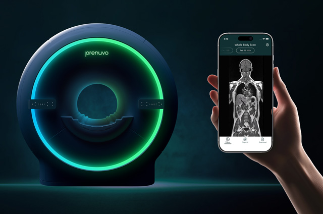
CT scans take longer than regular x-rays. You need to lie still on a table while they are being done. During the test, the table moves in and out of the scanner, a ring-shaped machine that completely surrounds the table. You might feel a bit confined by the ring you have to lie in when the pictures are being taken. CT scans are often used to diagnose pancreatic cancer and are helpful in staging the cancer (determining the extent of its spread). CT scans show the pancreas fairly clearly and often can confirm the location of the cancer. CT scans can also show the organs near the pancreas, as well as lymph nodes and distant organs where the cancer might have spread. The CT scan can help to determine whether surgery is a good treatment option.
CT scans can also be used to guide a biopsy needle precisely into a suspected area of spread. For this procedure, called a CT-guided needle biopsy, the patient remains on the CT scanning table as a radiologist advances a biopsy needle toward the location of the mass. CT scans are repeated until the doctors are sure that the needle is within the mass. A biopsy sample is then removed and looked at under a microscope.
- Magnetic resonance imaging (MRI) - MRI scans use radio waves and strong magnets instead of x-rays. The energy from the radio waves is absorbed by the body and then released in a pattern formed by the type of body tissue and by certain diseases. A computer translates the pattern into a detailed image of parts of the body. Not only does this produce cross-sectional slices of the body like a CT scanner, it also produces slices that are parallel with the length of the body. A contrast material might be injected just as with CT scans, this but is used less often.
Most doctors prefer CT scans to look at the pancreas, but an MRI may sometimes provide more information. MRI scans are also particularly helpful in looking at the brain and spinal cord.
MRI scans are a little more uncomfortable than CT scans. They take longer -- often up to an hour. You may have to lie inside a narrow tube, which is confining and can upset people with a fear of enclosed spaces. Newer, "open" MRI machines can help with this if needed. The MRI machine makes loud noises that you may find disturbing. Some places provide headphones with music to block this out.
- Positron emission tomography (PET) scan - PET scans involve injecting glucose (a form of sugar) that contains a radioactive atom into the blood. Because cancer cells in the body are growing rapidly, they absorb more of the radioactive sugar than the normal cells. A special camera can then create a picture of areas of radioactivity in the body. The picture is not finely detailed like a CT or MRI scan, but it provides helpful information. This test is useful to see if the cancer has spread to lymph nodes. PET scans are also useful when your doctor thinks the cancer has spread, but doesn't know to where.
PET/CT scans combine a CT scan and a PET scan to even better pinpoint the tumor. This test may be especially useful for spotting cancer that has spread beyond the pancreas and wouldn't be treatable by surgery. It may be a useful test for staging the cancer. It may even be able to spot early cancers.
- Ultrasonography - Ultrasound uses sound waves to produce images of internal organs such as the pancreas. For an abdominal ultrasound, a wand-shaped probe called a transducer is placed on the skin of the abdomen. It emits sound waves and detects the echoes as they bounce off internal organs. The pattern of echoes is processed by a computer to produce an image on a screen.
The echoes made by most pancreatic tumors differ from those of normal pancreas tissue. Different echo patterns can help distinguish some types of pancreatic tumors from one another.
If signs and symptoms indicate that a pancreatic cancer is likely, a CT scan is often more useful than ultrasound for an accurate diagnosis. But if it's not clear whether certain other diseases may account for the patient's signs or symptoms, ultrasound may be done.
Endoscopic ultrasound is more accurate than abdominal ultrasound and is probably the best way to diagnose pancreatic cancer. This test is done with an ultrasound probe that is attached to an endoscope -- a thin, lighted, flexible, fiber optic tube that doctors use to look at the inside of the intestinal tract. Patients are first sedated (given medicine to make them sleepy). The probe is then passed through the mouth or nose, through the esophagus (also known as the "food pipe") and stomach, and into the first part of the small intestine. The probe can then be pointed toward the pancreas, which sits next to the small intestine. The probe is on the tip of the endoscope, so it can get very close to the area where the tumor is to take pictures. This is a very good way to look at the pancreas. It is better than CT scans for spotting small tumors. If a tumor is seen, it can be biopsied during this procedure.
- Endoscopic retrograde cholangiopancreatography (ERCP) - For this procedure, an endoscope (a thin, lighted, flexible tube) is passed down the patient's throat, through the esophagus and stomach, and into the first part of the small intestine. The doctor can see through the endoscope to find the ampulla of Vater (the place where the common bile duct is connected to the small intestine). The doctor guides a catheter (a very small tube) from the end of the endoscope into the common bile duct. A small amount of dye (contrast material) is then injected through the tube into the common bile duct and x-rays are taken. This dye helps outline the bile duct and pancreatic duct. The x-ray images can show narrowing or blockage of these ducts that might be due to pancreatic cancer. The doctor doing this test can also put a small brush through the tube to remove cells for a biopsy (to view under a microscope to see whether or not they look like cancer). This procedure is usually done while the patient is sedated.
ERCP can also be used to place a stent (small tube) into the bile duct to keep it open if a nearby tumor is pressing on it. This is described in more detail in the section on palliative surgery.
- Angiography - This is an x-ray procedure for looking at blood vessels. A small amount of contrast material is injected into an artery to outline the blood vessels. After this, x-rays are taken. Angiography can show whether blood flow in a particular area is blocked or compressed by a tumor. It can also show any abnormal blood vessels (feeding the cancer) in the area. This test can be useful in finding out if a pancreatic cancer may have grown through the walls of certain blood vessels. Mainly, it helps surgeons decide whether the cancer can be completely removed without damaging vital blood vessels and helps them plan the operation.
Angiography can be an uncomfortable procedure because the radiologist who performs it has to put a small catheter into the artery leading to the pancreas. Usually the catheter is put into an artery in your inner thigh and threaded up to the pancreas. A local anesthetic is often used to numb the area before inserting the catheter. Then the dye is injected quickly to outline all the vessels while the x-rays are being taken.
Blood tests - Several types of blood tests may be used to help diagnose pancreatic cancer or to help determine treatment options if it is found. Blood tests that look at levels of different kinds of bilirubin (a chemical made by the liver) are useful to decide whether a patient's jaundice is due to a disease of the liver or to blockage (by a gallstone, a tumor, or other disease) of bile flow. Elevated blood levels of the tumor markers CA 19-9 and carcinoembryonic antigen (CEA) may point to a diagnosis of pancreatic cancer, but these tests aren't always accurate.
Other blood tests can help evaluate a patient's general state of health (such as liver, kidney, and bone marrow function). These tests can also help determine whether they'll be able to withstand the stress of a major operation.
- Biopsy - A patient's history, physical exam, and imaging test results may strongly suggest pancreatic cancer, but the only way to be sure is to remove a small sample of tumor and look at it under the microscope. This procedure is called a biopsy.
There are several types of biopsies. The procedure used most often to diagnose pancreatic cancer is called a fine needle aspiration (FNA) biopsy. For this test, a doctor inserts a thin needle through the skin and into the pancreas. The doctor uses CT scan images or endoscopic ultrasonography to view the position of the needle and make sure that it is in the tumor.
Doctors can also biopsy the tumor by using the endoscopic ultrasound to place the needle directly through the wall of the duodenum into the tumor. In either case, small tissue samples can be removed through the needle. The main advantages of the test are that the patient does not require general anesthesia (is not "asleep") during the test, and major side effects are rare.
In the past, surgical biopsies were performed more commonly. This type of biopsy requires a laparotomy (a large incision through the skin into the wall of the abdomen to examine internal organs). Areas that look or feel abnormal can be sampled by removing a small portion of tissue with a scalpel or a needle. The surgeon may use a thin needle (as in a fine needle aspiration biopsy). More commonly, surgeons use a wider needle that removes a cylindrical core of tissue about 1/2 inch long and less than 1/8 inch in diameter (called a core needle biopsy). The main drawback of this type of biopsy is that the patient must have general anesthesia and remain in the hospital for a period of time to recover.
Laparotomy is now rarely recommended. Doctors prefer to use laparoscopy (sometimes called keyhole surgery) as a way of looking at and perhaps taking a piece of the pancreas with a biopsy. Patients are usually sedated for this procedure. The surgeon makes several small incisions in the abdomen and inserts small telescope-like instruments into the abdominal cavity. One of these is usually connected to a video monitor. The surgeon can view the abdomen and see how big the tumor is and whether it has spread, and may take tissue samples as well.
Most doctors who treat pancreatic cancer try to avoid surgery unless imaging tests suggest that an operation might be able to remove all of the visible cancer. Even after doing imaging tests and laparoscopy, there are times when the surgeon begins an operation with the intent of removing the cancer but finds during surgery that it has spread too far to be removed completely. In these cases, a sample of the cancer is taken only to confirm the diagnosis, and the rest of the planned operation is stopped.


